Heart & Vascular
Trusted Expertise in Cardiac Care
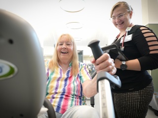
Heart and vascular specialists at University of Vermont Health are leading experts in the prevention and treatment of cardiovascular diseases. We offer full-spectrum cardiovascular services, including chronic disease management and heart surgery. Each year, we provide advanced care for thousands of patients throughout Vermont and northern New York.
Our cardiology specialists work as a team across our health system, so no matter where you live, you have access to compassionate and coordinated expertise.


Why Choose UVM Health?
- Expert physicians: Our team includes nationally recognized experts with advanced training and vast experience in heart and vascular diseases. As a health system anchored by an academic medical center, we are actively involved in training the next generation of physician leaders to propel the field forward.
- Coordinated care, accessible: At UVM Health, you’re always connected to the expertise of a much larger team of cardiologists, electrophysiologists, cardiovascular surgeons and other specialists who work together to give you seamless, personalized care. If you need more specialized treatment, we quickly coordinate transfers and referrals.
- Evidence-based, research-driven treatments: Many of our physicians are also active researchers that participate in and lead national clinical trials designed to improve the diagnosis and treatment of heart and vascular disease. Our involvement in research gives our patients access to promising emerging therapies that aren’t widely available.
- Advanced treatments: We perform high volumes of advanced heart and vascular procedures and percutaneous structural heart procedures, such as transcatheter aortic valve replacements (TAVRs).
Heart & Vascular Services
Conditions
Treatments & Tests
Advancing Medicine & Quality of Care

Better care, advanced procedures and cutting-edge treatments for you and your loved ones.

Improving the quality of your care now and into the future by training the next generation of health care professionals.

Immediate access to new treatments before they are widely available.