Heart Valve Disease
Heart Valve Disease Care at UVM Health
Heart valve disease, also called structural heart disease or valvular heart disease, occurs when the valves of the heart are not opening or closing properly. Your heart has four valves that work to keep blood flowing in the right direction. When one of the valves is not functioning correctly, blood cannot flow in and out of the heart as it should.
Heart valve disease is a common condition that can be congenital (appearing at birth) or acquired (developing over a period of time). Mild cases of heart valve disease may not cause symptoms or require treatment, but more serious cases will weaken the heart and can lead to heart failure.

Why Choose UVM Health?
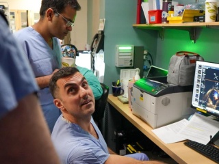
At University of Vermont Health, we take a coordinated, team-based approach to caring for patients with valvular disease. Our network of cardiology providers gives you access to advanced heart specialists, so no matter where you live in Vermont or northern New York, you can get personalized care that helps you get back to doing the things you love.
We offer:
- Specialized experts: Our structural heart team is made up of general cardiologists, interventional cardiologists, cardiothoracic surgeons, cardiac electrophysiologists, heart failure specialists and imaging specialists who collaborate to determine what treatment is right for you.
- 24/7 rapid response: Our Extracorporeal Membrane Oxygenation activation system is an advanced life-support treatment we can mobilize within an hour for critically ill patients.
- Advanced treatment options: When medications alone don’t manage heart valve disease, you have access to advanced interventional procedures to repair or replace the damaged valve, including leading-edge minimally invasive procedures. We are also the only providers in Vermont offering catheter-based treatments as an alternative to open-heart surgery.
- Care grounded in research: Our care is backed by research and we participate in national clinical trials to establish new treatment options, such as transcatheter aortic valve replacement (TAVR) and transcatheter edge-to-edge repair (TEER).
Heart Valve Disease Risk Factors
Valvular disease is associated with a number of underlying conditions, including:
- Connective tissue disease
- Endocarditis (an infection of the heart)
- Calcium deposits
- Coronary artery disease
- Heart failure
- Heart attack
- Congenital heart defects
- Rheumatic fever
- Enlarged aorta
- Trauma to the heart
Wellness & Prevention
Evidence shows that proactive health care focused on preventing illness leads to better outcomes. We're here to help you live a healthier, happier life. We offer wellness and prevention services to empower you to take control of your health.

Types of Heart Valve Disease
There are many types of heart valve disease, each affecting the heart’s valves in different ways.
This common form of heart valve disease occurs when the mitral valve doesn’t close properly. The mitral valve controls blood flow to the left side of your heart, letting blood flow in one direction from the upper chamber of the heart (atrium) to the lower chamber (ventricle). In a mitral valve prolapse, the valve lets blood flow through normally but bulges backwards after it has closed.
This type of heart valve disease is typically caused by endocarditis (an infection of the heart) or connective tissue diseases. It can also be passed down through family members.
In most cases, MVP does not cause heart damage and does not require treatment. However, it can increase your chances of developing mitral valve regurgitation.
When the mitral valve does not close properly, blood may leak or flow backwards (regurgitate) from the lower left heart chamber (ventricle) to the upper left chamber (atrium). This is called mitral valve regurgitation (MVR). In serious cases, this condition can lead to a backup of blood in the left atrium and lungs and damage the left ventricle, ultimately leading to heart failure.
There are two types of MVR. Primary mitral valve regurgitation is caused by a structural problem with the valve itself, often caused by a congenital heart defect, mitral valve prolapse or calcium buildup in the mitral valve. Primary MVR is treated surgically, by replacing the defective parts of the valve.
Secondary mitral valve regurgitation occurs when another condition (such as a heart attack or coronary artery disease) causes the valve to leak. Secondary MVR is addressed by first treating the underlying condition, then repairing the mitral valve after your medications are optimized.
MVR does not always cause symptoms and may never require treatment. In these cases, your provider may recommend ongoing monitoring to make sure the leak does not get worse and prescribe medication to help your blood flow more easily.
Mitral valve stenosis is a narrowing of the mitral valve, preventing it from opening completely. This causes pressure to build up in the left atrium and the lungs, resulting in shortness of breath. It also prevents the left ventricle from filling up with blood, making it harder for people to exercise. As stenosis progresses, the valve gets narrower, eventually leading to shortness of breath even at rest.
Mitral valve stenosis is caused by damage to the valve, which can be a result of calcium buildup, congenital heart defects or rheumatic fever.
Mild or moderate cases may not cause symptoms, and can be managed through long-term monitoring, medication and lifestyle changes. More serious cases usually require a procedure to dilate or replace the valve, such as balloon valvuloplasty, in which a balloon is inserted into the valve via catheter and inflated to improve blood flow.
The aortic valve works like a one-way gate, controlling blood flow from the heart to the aortic valve and the rest of the body. When it does not close properly, some of the blood can leak backwards (regurgitate) into the left ventricle. This means the body does not get enough blood, and the heart must work harder to make up for it.
Aortic valve regurgitation can be long-term (chronic) or come on suddenly (acute) and is usually a result of damage to the valve. Common causes of chronic aortic valve regurgitation include congenital heart defects, calcium buildup in the valve or an enlarged aorta. You can have chronic aortic valve regurgitation for years and not experience symptoms.
Acute aortic valve regurgitation is often caused by endocarditis or trauma to the heart and requires immediate medical attention.
Aortic valve stenosis is a narrowing of the aortic valve, preventing it from opening completely. This forces the heart to work harder to pump blood through the valve, causing pressure to build up in the ventricle and thickening the heart muscle. As stenosis progresses, the valve gets narrower, eventually leading to heart failure.
Aortic valve stenosis is caused by damage to the valve, which can be a result of calcium buildup on the valve, congenital heart defects or rheumatic fever.
Mild or moderate cases may not cause symptoms, and can be managed through long-term monitoring, medication and lifestyle changes. More serious cases usually require surgery to fix or replace the valve.
When the tricuspid valve does not close properly, blood may leak or flow backwards (regurgitate) from the lower right heart chamber (ventricle) to the upper right chamber (atrium). This is called tricuspid valve regurgitation (TVR). In serious cases, this condition can lead to a backup of blood in the right atrium and veins, ultimately leading to right-sided heart failure. Symptoms include leg swelling, liver congestion and abdominal distension.
TVR does not always cause symptoms and may never require treatment. In these cases, your provider may recommend ongoing monitoring and prescribe diuretics to control the congestion and swelling. There are catheter-based options that can be considered for repairing the tricuspid valve or minimizing the congestion.
Symptoms of Heart Valve Disease
In many cases, heart valve disease does not cause symptoms. You may not know you have the condition, and can live with it for many years before it requires treatment. As the disease progresses, symptoms may appear.
Common signs and symptoms of valvular disease can include:
- Shortness of breath, especially with activity
- Fatigue
- Heart murmur
- Dizziness
- Fainting
- Abnormal heartbeat (arrhythmia)
- Chest pain or pressure
- Trouble breathing at night
- Swelling in the legs and sometimes the rest of the body (edema)
Thankfully, there is a minimally invasive procedure called TAVR that could fix my heart issue and I avoided open-heart surgery.
Diagnosing Heart Valve Disease
If your doctor suspects you have heart valve disease, they will discuss any symptoms with you and ask questions about your medical history and family history. They will then conduct a thorough physical exam and listen to your heart to see if they can detect a murmur. Based on their findings, they will order further tests to confirm the diagnosis.
Heart valve diagnosis and testing may include:
- Echocardiogram: Creates a video image of your heart pumping blood and determine what type of heart defect you have
- Positron emission tomography (PET): Studies how well your organs and tissues are working
- Cardiac magnetic resonance imaging (MRI): Evaluates the structure and function of the heart and other organs in the chest cavity, including blood vessels
- Cardiac catheterization and biopsy: Produces a detailed picture of your heart structure and valve function using a long, thin tube (catheter), a special dye and imaging tools
Heart Valve Disease Treatment
There are a number of options when it comes to treating valvular disease. The course of treatment your cardiologist recommends will depend on the type and severity of your disease. Heart valve disease treatment options include:
In order to protect your heart from further damage and to avoid serious conditions such as endocarditis and heart failure, your cardiologist may recommend the following protective measures and lifestyle changes:
- Communicate your condition to your doctors, your dentist, and your friends and family. You can also obtain an identification card from the American Heart Association with this information in the event of a medical emergency.
- Treat infections promptly, under the guidance of your medical provider. Stay up to date with your flu and COVID-19 vaccinations.
- Maintain oral hygiene to avoid infections in your teeth and gums. Make regular appointments with your dentist.
- Take antibiotics before you undergo any procedure that may cause bleeding.
- Eat a heart-healthy diet. Limit sodium, sugar and alcohol.
- Stay active. Your doctor can advise the safest type and level of exercise for you.
- Maintain a healthy weight. Lose weight if necessary.
- Do not smoke. If you need help, talk to your doctor about smoking cessation programs.
- Manage your health conditions. Stay on top of other conditions such as diabetes, high blood pressure and high cholesterol.
Medications may be prescribed alone or in combination with other heart valve disease treatments. Your cardiologist may prescribe one or more of the following:
- Angiotensin-converting enzyme (ACE) inhibitors: Reduces angiotensin which is a substance that causes blood vessels to constrict and increases blood pressure
- Antiarrhythmics: Controls abnormal heart rhythms
- Anticoagulants (blood thinners): Dissolve blood clots or prevent them from forming
- Beta blockers: Helps your heart relax, slow your heart rate and lower your blood pressure
- Diuretics: Also known as “water pills,” they are used to reduce swelling and fluid buildup
- Vasodilators: Encourage blood to flow forward rather than backward
Depending on your diagnosis and the type of heart valve disease you have, your cardiologist may recommend a heart valve repair or replacement. Interventional cardiologists and cardiothoracic surgeons at UVM Health offer the following minimally invasive heart surgeries to treat valvular disease:
- Minimally invasive aortic valve surgery: Procedure to replace your damaged aortic valve with a new valve
- Minimally invasive mitral valve surgery: Procedure to reinforce your mitral valve, widen the valve, repair holes in the valve or reconnect valve flaps
- Minimally invasive tricuspid valve surgery: Catheter-based procedures reduce tricuspid valve leakage by partially closing the valve with a clip or replacing the valve through the blood vessel.
- Minimally invasive thoracic aortic aneurysm surgery: Procedure to strengthen your weakened aorta and prevent an aneurysm from rupturing
Extracorporeal Membrane Oxygenation (ECMO) is an advanced life-support treatment that temporarily takes over the work of your heart and lungs. Using a specialized machine, ECMO oxygenates your blood and returns it to your body — giving your organs time to heal.
Our multidisciplinary team includes specialists in cardiac surgery, interventional cardiology, cardiovascular ICU, pulmonary critical care, ICU nursing, respiratory therapy, rehabilitation and perfusion.
Locations Near You
Share your location to see nearby providers and availability
62 Tilley Drive
Suite 101
South Burlington, VT 05403
118 Tilley Drive
Suite 102
South Burlington, VT 05403-4450
115 Porter Drive
Middlebury, VT 05753
75 Park Street
Elizabethtown, NY 12932
101 Adirondack Drive
Suite 1
Ticonderoga, NY 12883
78 Park Street
Elizabethtown, NY 12932
133 Park Street
Malone, NY 12953
130 Fisher Road
Berlin, VT 05602
210 Cornelia Street
Ste 104
Plattsburgh, NY 12901
62 Tilley Drive
Suite 101
South Burlington, VT 05403-4407