Cardiomyopathy
Cardiomyopathy Care at UVM Health
Cardiomyopathy is a condition that adversely affects the performance of your heart muscle, making it harder for your heart to pump blood and deliver it to the rest of the body. The disease can be acquired (as a result of heart damage from another condition) or genetic (passed down from a parent).
Cardiomyopathy may not cause symptoms in the early stages of the disease, but left untreated, it can lead to the development of symptoms associated with heart failure. Treatment depends on the type of cardiomyopathy you have and may involve a combination of both non-surgical and surgical methods.
Why Choose UVM Health?
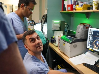
At University of Vermont Health, we take a coordinated, team-based approach to caring for patients with cardiomyopathy. Our network of cardiology specialists gives you access to advanced heart specialists, so no matter where you live in Vermont or northern New York, you can get personalized care that helps you get back to doing the things you love.
We offer:
- Specialized experts: Your treatment team may include advanced heart failure specialists, interventional cardiologists, cardiac surgeons, cardiac electrophysiologists and imaging specialists. These physicians collaborate to determine which treatment is right for you.
- Advanced treatment options: Interventional cardiologists at UVM Health are the only specialists in Vermont offering catheter-based treatments in patients with specific conditions as an alternative to open-heart surgery.
- Care grounded in research: We offer one of the top cardiology programs in the region, giving you access to leading-edge technology and research-backed treatments, including promising but yet unproven new therapies through clinical trials.
Types of Cardiomyopathy
There are three main types of cardiomyopathy, each caused by different factors. In general, cardiomyopathy occurs as a result of damage to the heart, such as from a heart attack, or a person may have a genetic predisposition for developing the condition.
Dilated cardiomyopathy, or heart failure reduced ejection fraction, is one of the more common types of cardiomyopathy. It can develop after a heart attack or other conditions that damage the heart, such as a viral illness. In this condition, your heart’s ability to pump blood is reduced, resulting in the heart ejecting less blood with each beat. Over time, your heart may dilate (stretch), and you may develop symptoms of heart failure, including shortness of breath, fatigue, weight gain and leg swelling.
Although this form of cardiomyopathy can affect people of all ages, it occurs most often in middle-aged or older people, and is more likely to affect men. Some people may have a family history of the condition.
Restrictive cardiomyopathy, or heart failure preserved ejection fraction, causes the heart muscle to become stiff, meaning the heart can’t stretch enough for the heart chambers to fill properly. Blood then backs up in the circulatory system, which can lead to congestive heart failure.
Restrictive cardiomyopathy can happen at any age, but it most often affects older people. The condition may be caused by diseases that affect the heart, such as hypertension, diabetes and obesity, or it may occur with no known cause (idiopathic cardiomyopathy). Approximately 50% of patients with heart failure have restrictive cardiomyopathy.
This type of cardiomyopathy happens when the heart muscle grows too thick, causing the heart to get bigger and its chambers to get smaller. This thickening muscle stiffens the heart and affects its performance.
Hypertrophic cardiomyopathy can begin at any age, but the condition tends to be more serious the earlier in life it starts. Hypertrophic cardiomyopathy runs in families and is the most common genetic heart disease.
Symptoms of Cardiomyopathy
You may not experience cardiomyopathy symptoms in the early stages of the condition, but as it advances, symptoms are likely to appear. Regardless of the type of cardiomyopathy you have, symptoms tend to get worse without treatment. This process can happen slowly or quickly.
Common signs and symptoms of cardiomyopathy can include:
- Shortness of breath, especially with activity
- Fatigue
- Difficulty breathing when lying down
- Swelling in your legs (edema)
- Chest pain
- A fast or irregular heartbeat (arrhythmia)
Diagnosing Cardiomyopathy
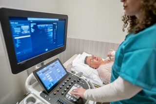
If your doctor suspects you may have cardiomyopathy, they will discuss your symptoms with you and conduct a thorough physical exam. They will then refer you to a cardiovascular specialist, who will use a full range of cardiac imaging and stress testing to diagnose your condition accurately and efficiently.
Cardiomyopathy diagnosis and testing may include:
- Echocardiogram: Creates a video image of your heart pumping blood and determine what type of heart defect you have
- Electrocardiogram (EKG): Records your heart’s electrical activity and identify any abnormal rhythms and stress to the heart
- Cardiac magnetic resonance imaging (MRI): Evaluates the structure and function of the heart and other organs in the chest cavity, including blood vessels
- Cardiac stress testing: Evaluates you for coronary artery disease. This may include echo or nuclear perfusion to image your heart and involves either an exercise treadmill test or medication that simulates the effects of exercise.
- Cardiac catheterization: Identifies blocked or narrowed arteries using a long, thin tube (catheter), a special dye and X-ray cameras to take images of the arteries that feed your heart muscle
- Blood tests: Look for chemical markers of cardiomyopathy or other diseases that can lead to heart failure
- Chest X-ray: Fully examines your heart, lungs, airway, blood vessels and lymph nodes
Cardiomyopathy Treatment
The goals of cardiomyopathy treatment are to manage your symptoms, prevent your condition from worsening and reduce your risk of complications.
The course of treatment your cardiologist recommends will depend on the results of your diagnostic testing and the type of cardiomyopathy you have, among other factors.
Cardiomyopathy treatment options include:
Medications may be prescribed alone or in combination with other cardiomyopathy treatments. Your cardiologist may prescribe one or more of the following:
- Angiotensin-converting enzyme (ACE) inhibitors: Reduce angiotensin, a substance that causes blood vessels to constrict and increases blood pressure
- Angiotensin receptor blockers (ARBs): Block the action of angiotensin
- Sacubatril/Valsatan (Entresto): Improve your heart’s ability to pump blood
- Beta blockers: Help your heart relax, slow your heart rate and lower your blood pressure
- Digitalis (digoxin): Help your heart beat more strongly
- Diuretics: Also known as “water pills,” reduce swelling and fluid buildup
- SGLT2 inhibitors: Lower your blood sugar levels
Your cardiologist may determine that your heart needs assistance from a medical device. Options for surgically implanted devices include:
- A pacemaker: Helps your heart maintain a regular heartbeat
- Implantable cardioverter-defibrillator (ICD): A special type of pacemaker that monitors your heart rate and deliver electric shocks to the heart when necessary
Cardiac catheterization uses thin, flexible tubes (catheters) inserted into an artery in your groin to access the heart. This procedure helps patients avoid major surgery and a lengthy recovery process.
The cardiac catheterization procedure for cardiomyopathy is called alcohol septal ablation. When the area that divides the right and left chambers of the heart (septum) becomes too thick, the lower left heart chamber (left ventricle) gets blocked and is unable to pump normally.
The thickened septum can be reduced in size by injecting alcohol through a catheter to destroy some of the overgrown heart muscle. This decreases the blockage and improves the left ventricle’s pumping ability.
Locations Near You
Share your location to see nearby providers and availability
62 Tilley Drive
Suite 101
South Burlington, VT 05403
118 Tilley Drive
Suite 102
South Burlington, VT 05403-4450
115 Porter Drive
Middlebury, VT 05753
75 Park Street
Elizabethtown, NY 12932
101 Adirondack Drive
Suite 1
Ticonderoga, NY 12883
78 Park Street
Elizabethtown, NY 12932
133 Park Street
Malone, NY 12953
130 Fisher Road
Berlin, VT 05602
210 Cornelia Street
Ste 104
Plattsburgh, NY 12901
62 Tilley Drive
Suite 101
South Burlington, VT 05403-4407