Photo of the entrance to UVM Medical Center's facility on Tilley Drive in South Burlington.

Cardiology - Tilley Drive

 (802) 847-2533

62 Tilley Drive
Suite 101
South Burlington, VT 05403-4407

Monday: 8:00 AM - 5:00 PM
Tuesday: 8:00 AM - 5:00 PM
Wednesday: 8:00 AM - 5:00 PM
Thursday: 8:00 AM - 5:00 PM
Friday: 8:00 AM - 5:00 PM

Echocardiography: What You Need to Know

Echocardiography is a powerful, non-invasive diagnostic tool that can be applied at the bedside. Its effectiveness requires a thorough understanding of both the technology that enables image acquisition and the clinical implications of the images. The University Cardiology Associates offer patients a hospital based laboratory with state of the art echocardiography equipment.

Images are acquired by highly skilled sonographers and interpreted by cardiologists with specific subspecialty training in echocardiography. The transportability of echocardiography allows us to offer these services to patients at our outpatient facilities on Tilley Drive in South Burlington as well as at area hospitals.The laboratory is directed by Dr. Marc Tischler, a national authority, and is staffed by residents, students, and at least two fellows in training.

Dr. Tischler is a hands-on director who spends his days in the laboratory, interpreting studies as well as teaching and mentoring residents and fellows. Because the laboratory is focused on research, teaching, and direct patient care, the attending faculty bring a depth of insight to interpretation of results that directly translates into improved clinical diagnosis, patient care, and outcomes.

Echocardiography Procedures and Treatments

The most commonly performed echocardiogram is the transthoracic echocardiogram , that is acquired by placing the probe on the outside of the chest wall with a gel-like substance to transmit sound waves into the body.

The Doppler echocardiogram evaluates blood flow in the heart and blood vessels. This procedure measures the speed and direction of the blood flow within the heart. It screens the four valves for leaks or other abnormalities. With Doppler echocardiograms, as the transducer moves over your heart you will hear a "whooshing" sound much like that of a washing machine. This sound reflects the movement of blood in your heart.

The stress echocardiogram combines an echocardiographic examination with a treadmill or bike exercise test, or medication that simulates the effect of exercise on the heart. Both forms of stress echocardiograms can be used to diagnose the presence and extent of narrowing of the coronary arteries as well as the significance of valvular heart disease.

The contrast echocardiogram combines an echocardiogram with an administration, through a vein, of a sterile contrast solution, that improves visualization of the heart. This is a harmless agent that has no significant side effects. You will need to have an IV started to receive a contrast echocardiogram.

The Transesophageal echocardiogram entails placing a miniature ultrasound probe into the esophagus, or food pipe, behind the heart. This allows the physician to obtain superior resolution of cardiac structure and function. Transesophageal echocardiograms are typically performed to evaluate strokes and transient ischemic attacks (TIA), complex valvular heart disease including previous valve replacements congenital heart disease, pathology of the aorta, and whenever superior resolution is needed.

What is an echocardiogram?

An echocardiogram is a noninvasive test that uses sound waves to look at the structure and function of your heart. A transducer, which is a medical device, converts electrical energy into sound waves that are then directed into your body. When the sound waves reach your heart, they are reflected back to the body surface where they are received by the transducer and electronically converted into a picture.

The transducer is moved back and forth on the skin, but there is no unpleasant sensation. A gel, similar to mineral oil, is used to keep the transducer in close contact with the skin.

What are the risks?

This is a safe, painless test. It uses the same techniques as the ultrasound test done during pregnancy. The echocardiogram shows a much clearer and more detailed image of your heart. The dynamic nature of the examination provides more information about function than is possible with an x-ray study.

Cardiac Ultrasound Basic Methods

An echocardiogram is a non-invasive ultrasound evaluation of the heart. Cardiac ultrasound, otherwise known as echocardiography, utilizes the same principles of sound wave investigation as used for obstetric and gynecologic evaluations of pregnant women, evaluation of the gall bladder and other abdominal structures. The basic principle involves directing ultrasound waves into the body and then analyzing the reflected wave.

The reflected wave is converted into an actual image of the heart and displayed in a real time or motion format. The ultrasound is directed out of a transducer which is a hand held device that can be moved over the chest to image the heart from different positions.

There are no side-effects to the use of ultrasound for diagnostic purposes. This pertains to side-effects experienced by the patient, pregnant women, fetus or the operator. To obtain better contact between the transducer and the skin, an ultrasound gel is applied to the chest wall. This has the consistency of thick mineral oil and is not associated with any skin irritation or allergy. Additionally, so that heart events can be timed, three EKG leads are placed on the chest.

Doppler ultrasound utilizes the same equipment that are used to create an actual picture of the heart. Doppler ultrasound relies on the same principle used by police to detect the speed of your automobile with radar. By analyzing the ultrasound wave that comes back from the heart in a slightly different manner, the ultrasound instrument can determine the direction and speed at which blood flows through your heart.

This can be utilized to determine the severity of a leaking valve or a valve that is stenosed (too tight) and also to identify intracardiac shunts, a so called "hole in the heart". The combination of routine two-dimensional ultrasound and Doppler ultrasound facilitates a rapid, painless, safe, and complete evaluation of the anatomy of the heart as well as its function.

Transthoracic echocardiography is the most common type of cardiac ultrasound utilized. At The University of Vermont Medical Center, approximately 6,500 patients undergo transthoracic echocardiography each year. The procedure requires from 15 to 60 minutes depending on the complexity of the examination and is accomplished without sedation or special patient preparation.

It is not necessary to withhold medications or to abstain from eating before the test. Transthoracic echocardiograms are performed in The UVM Medical Center Echocardiography Laboratory and on a portable basis in the Emergency Department and the Intensive Care Units. In addition echocardiograms are performed in our out patient facility on Dorset Street as well as outreach programs in St. Albans, VT, Massena, NY, and Ticonderoga, NY.

The procedure for recording a transthoracic echocardiogram involves having the patient disrobe from the waist up. Hospital gowns are provided. Three electrocardiographic leads are then applied to the chest. Typically the best images will be obtained with the patient lying in a left lateral decubitus position, that is on the left side.

A pillow or wedge will be provided to allow a relaxed body position. The sonographer then applies echocardiographic gel to the area of the chest immediately adjacent to the breast bone and also to the area just under the left breast. The transducer is then gently maneuvered over the chest to provide the different views of the heart.

When the Doppler aspects of the ultrasound instrument are activated you will actually hear the sound of blood moving through the heart. For other views you may be asked to lie on your back with your knees bent, or occasionally to sit in an upright position so that the transducer can be placed in the area just above the breast bone in your neck. The entire examination usually requires 30 minutes. When the procedure is finished the ECG leads will be removed and you will be given a towel to wipe-off the gel. The gel is clear, odorless, non-irritating, and does not stain clothing.

The ultrasound examination will be interpreted by one of our faculty echocardiographers. For hospitalized patients results are available within a two to three hour period. Your referring physician will receive the final report by mail or facsimile, within 24 hours. Our computer network allows access to reports online.

Information that is available from routine transthoracic echocardiography includes an assessment of the size, shape and function of your heart, including identification of areas that may have been damaged by infection or heart attack. Additionally, the echocardiogram is an ideal tool for determining the function of the four cardiac valves and detecting the presence and severity of damage to the valves that may be caused by rheumatic fever or infection. Other conditions such as pericardial fluid (fluid in sac surrounding the heart) or congenital heart defects such as atrial septal defects (hole in the heart) are accurately detected and characterized as well.

Treadmill Stress Echocardiograms

Stress echocardiograms involve performing transthoracic echocardiography before and after cardiovascular stress. Stress echocardiograms are commonly used for detection of coronary artery disease, or in determining the impact that a heart attack may have had on cardiovascular function. Stress echocardiograms are less commonly performed to evaluate pulmonary artery pressures in patients with lung disease or pulmonary hypertension and to evaluate the significance and progression of valvular heart disease.

The ultrasound portion of a stress echocardiogram confers no risk or discomfort to the patient. There is a small risk to the patient that is associated with the stress component of the test, but this risk is not increased by the addition of ultrasound. In many instances addition of ultrasound may confer an added margin of safety.

An exercise echocardiogram involves transthoracic echocardiography (see above) performed at baseline while you are resting and immediately after or during physical exercise. The physical exercise that is utilized may involve treadmill testing, in which case echocardiographic images are obtained before and immediately after exercise.

Transthoracic echocardiograms are recorded on videotape for future analysis. Stress echocardiograms similarly recorded on videotape but also captured in a digital format that allows the interpreter to view rest and stress images side by side. This facilitates the detection of new wall motion abnormalities.

Exercise or medications increase the extent to which the heart contracts. If a patient has had a myocardial infarction (heart attack) there will be an area of the heart muscle that does not contract normally at rest. This area may exhibit decreased contraction with stress. Those without a previous heart attack but blockage in their coronary artery may exhibit normal function at rest.

During stress an area of the heart that is deprived of contract effectively and becomes abnormal. By comparing the rest and exercise portions of the stress echocardiogram an experienced echocardiographer can determine the presence and severity of a coronary artery blockage.

Dobutamin Stress Echocardiography

Dobutamine stress echocardiography is a method to induce cardiac stress with a medication. To obtain accurate information from a stress test patients must have a reasonable exercise capacity. Approximately 30% of patients are in exercise capacity to exercise to an a extent sufficient that reveals coronary artery blockages. The limitation may be due to other medical illnesses such as diabetes, lung disease, peripheral vascular disease or even physical deconditioning.

In such cases, the heart can be stressed by infusing a medicine called dobutamine. Dobutamine is commonly used as an agent to treat patients in the hospital with low blood pressure and to improve circulation. For the past ten years it has also been used as a diagnostic testing agent. Dobutamine increases the heart rate and blood pressure and also the extent to which the heart contracts. It therefore mimics the effects of exercise.

To perform a dobutamine stress echocardiogram a conventional transthoracic echocardiogram is performed initially. An intravenous line is placed and dobutamine is infused initially at a low dose and then incrementally greater doses are administered every three minutes to increase the heart rate and overall cardiac work. During each of the three minute stages the heart is imaged using transthoracic echocardiography.

During infusion of the drug you can expect to feel your heart rate and the forcefulness of the heart contractions increase. This occasionally causes a disconcerting sensation because the patient is lying quietly at rest while the heart feels as if it is running a race. This is to be expected during infusion of dobutamine and does not represent any harmful effect. When one test is completed the infusion is discontinued and the effects resolve in five to ten minutes.

During the test your electrocardiogram will be continuously recorded and your blood pressure will be determined every three minutes. The dobutamine stress echocardiogram is typically performed by one of the cardiac sonographers and a Registered Nurse with specific training in cardiac stress testing. Faculty physicians are in the immediate vicinity as well. As for all types of cardiovascular stress testing, all equipment and medicines necessary to treat complications are available in the stress testing room.

As with exercise echocardiography the images are recorded on videotape and also "captured" in a digital format. This allows the interpreter to compare the function of the heart at rest with that at each stage of the dobutamine infusion.

Bicycle Stress Echocardiography

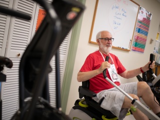
In selected cases bicycle exercise echocardiography will be used instead of treadmill exercise. Bicycle exercise is particularly useful for patients with valvular heart disease or for evaluated pulmonary artery pressures. It is also useful when the heart performance at each stage of exercise needs to be assessed. Bicycle exercise echocardiography confers no additional risk beyond that of treadmill exercise. It offers the advantage of imaging during exercise.

Why perform an exerciseechocardiogram?

Not all patients require exercise echocardiography. In many instances routine exercise treadmill testing with monitoring of the electrocardiogram (ECG) suffices for detection and characterization of coronary artery disease. Unfortunately, there are a number of patients in whom routine ECG monitoring is suboptimal.

Patients who have underlying abnormalities of the electrocardiogram such as a bundle branch block or left ventricular hypertrophy (thick heart muscle, commonly associated with high blood pressure) often have an inaccurate ECG response to exercise. For reasons that remain poorly understood many female patients, especially above the age of 40, the diagnostic accuracy when the ECG alone is utilized is limited.

Transesophageal Echocardiography

Transesophageal echocardiography is a specialized echocardiographic procedure performed in The UVM Medical Center Echocardiography Laboratory. At The UVM Medical Center, approximately 500 patients undergo transesophageal echocardiography each year. For this procedure the ultrasound transducer has been miniaturized so that the crystals creating the ultrasound energy are approximately the size of a fingernail. This crystal array is then mounted on a gastroscope that is virtually identical to the type of instrument a gastroenterologist (stomach doctor) would use to look for ulcers or other disease in the GI system.

A transesophageal echocardiogram is more complex than transthoracic or stress echocardiography. To perform a transesophageal echocardiogram the patient must swallow the endoscopic probe. Because this procedure can be associated with discomfort is performed after the patient has been sedated. The medicines used for sedation are identical to those used by a gastroenterologist when performing this procedure and may include combinations of Demerol, Versed, Fentanyl or other sedatives and narcotics.

The procedure is performed under what is called "conscious sedation" that implies a level of sedation the patient can still respond to instructions and is capable of conversation after the procedure. A topical anesthetic is sprayed into the throat to limit discomfort.

One side-effect of the sprays used for topical anesthesia is that they may induce a sensation that the throat is swelling, although no actual swelling occurs. The entire mouth, throat, and tongue will be numb and taste and temperature sensation diminished for a three to four hour period.

Additionally, the medicines used for conscious sedation impair judgment and reflexes and cause a variable degrees of drowsiness. For this reason patients are monitored by a Registered Nurse in the echocardiography laboratory until they have returned to near baseline. Outpatients must have a family member or other responsible adult to provide transportation home. You should not make important decisions, operate dangerous equipment or drive for eighteen hours after the procedure.

Because of the need to place the probe into the mouth and esophagus patients may occasionally experience mild throat discomfort for a day after the procedure. The entire procedure requires approximately 90 minutes to complete. The majority of this time will encompass explanation of explaining the procedure, obtaining informed consent, starting an IV for administration of medicine and observation following the procedure. The probe is in place for an average of 15 minutes.

Why Perform a Transesophageal Echocardiogram?

Transesophageal echocardiography provides a superior resolution of many structures of the heart not attained with transthoracic echocardiography and in some instances not attained with any other diagnostic test. Anatomically, the esophagus connects your mouth down with your stomach. In doing so it is adjacent to your aorta (the main blood vessel leading from the heart) and the left atrium of the heart. Because of the proximity of and lack of interference from the chest wall or lung, the resolution of the pictures obtained is substantially greater than that from transthoracic echocardiography.

Additionally, because the probe is behind the left atrium of the heart the image of the mitral valve is enhanced. A common indication for performing a transesophageal echocardiogram is to look for masses or clots in the heart. Because the ultrasound probe is now separated from the heart only by the wall of the esophagus, the accuracy for finding a clot inside the heart is increased dramatically.

Specific times in which a transesophageal echocardiogram is necessary and cannot be replaced by other tests include, evaluation of artificial mitral valves, looking for clots inside the left atrium or other causes for a patient to have had a stroke, evaluation of the aorta which may have been damaged by atherosclerotic disease or a tear in its lining (aortic dissection) or infections in the heart.

Contrast Echocardiography

Contrast echocardiography refers to the injection of an ultrasound contrast agent into the blood stream. Ultrasound contrast agents typically are composed of a solution of microbubbles or occasionally air filled microspheres. Current agents include simple agitation of sterile saline (salt water) that is frequently used to detect shunts inside the heart.

This type of contrast echocardiography is also used to enhance Doppler signals. There are several new contrast agents that have been approved for use in patients detection of cardiac function. Future developments in contrast echocardiography include development of contrast agents that may allow doctors to track the flow of blood into the myocardium (i.e. myocardial perfusion) using ultrasound.

Intravascular Ultrasound

Intravascular ultrasound is a technique in which the ultrasound transducer has been miniaturized to an even greater extent and is contained in the tip of a catheter that can be used in cardiac catheterization (see section on cardiac catheterization). This technique is used by physicians in the catheterization laboratory rather than in the echocardiography laboratory and allows them to characterize the walls of blood vessels that supply the heart. This evaluation is distinct from the complementary to that is obtained with contrast and x-rays.

Aderonke O. Adeniyi, MD
Cardiovascular Disease
Daniel D. Correa de Sa, MD
Clinical Cardiac Electrophysiology
Cardiovascular Disease
Harold L. Dauerman, MD
Interventional Cardiology
Cardiovascular Disease
Gregory L. Ehle, PA-C
Cardiovascular Disease
      	        
	  	  Catherine  Falduto, NP
Catherine Falduto, NP
Cardiovascular Disease
Eric A. Gauthier, MD
Interventional Cardiology
Cardiovascular Disease
Prospero B. Gogo, MD
Interventional Cardiology
Cardiovascular Disease
Susan A. Hamlyn-Prescott, NP
Cardiovascular Disease
William E. Hopkins, MD
Cardiovascular Disease
Roger G. Ishac, MD
Interventional Cardiology
Cardiovascular Disease
Friederike K. Keating, MD
Cardiovascular Disease
Rony N. Lahoud, MD
Interventional Cardiology
Cardiovascular Disease
Robert M. Lobel, MD
Clinical Cardiac Electrophysiology
Cardiovascular Disease
Daniel L. Lustgarten, MD, PhD
Clinical Cardiac Electrophysiology
Cardiovascular Disease
Margaret A. MacDonald, NP
Cardiovascular Disease
Richard L. Page, MD
Cardiovascular Disease
David J. Schneider, MD
Cardiovascular Disease
Nancy L. Strong, NP
Cardiovascular Disease
      	        
	  	  Nathaniel C. Thompson, MD
Nathaniel C. Thompson, MD
Clinical Cardiac Electrophysiology
Cardiovascular Disease
Peter C. Van Buren, MD
Advanced Heart Failure and Transplant Cardiology
Cardiovascular Disease
Matthew W. Watkins, MD
Cardiovascular Disease
Pierre Znojkiewicz, MD
Clinical Cardiac Electrophysiology
Cardiovascular Disease