Nuclear Medicine
Nuclear Medicine at UVM Health
A radiology specialty, nuclear medicine uses injected or ingested radioactive substances called “tracers” and a variety of imaging technology to create 3D pictures of the inside of your body. Nuclear medicine provides safe and effective procedures for diagnosing disease and seeing how well your organs and other body systems are working.
As a health system anchored by an academic medical center, we offer the region’s most comprehensive nuclear medicine program — from state-of-the-art equipment to advanced cancer therapies.
Why Choose UVM Health?
We offer:
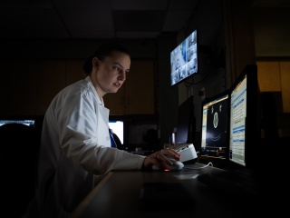
- Exceptional imaging capabilities: We perform all procedures using top-of-the-line digital imaging equipment with the highest picture resolution available to ensure accurate test results. No other health provider in our service area matches the technology we use to provide you with a safe, thorough and comfortable exam experience.
- Wide range of services: Our program offers you easy access to the full spectrum of nuclear medicine procedures, including PET scans, SPECT scans, bone scans, cardiac scans and hepatobiliary scans. In addition to imaging, we provide leading-edge nuclear medicine therapies, such as recently approved drugs Lutathera for the treatment of advanced neuroendocrine cancers and Pluvicto for the treatment of prostate cancer.
- Multidisciplinary approach: Our nuclear medicine specialists collaborate with your cardiologist, oncologist and other specialists, as well as your primary care physician, to personalize your care by determining the nuclear medicine options that will deliver the best results for your specific condition.
Conditions We Treat with Nuclear Medicine Procedures
Nuclear medicine imaging primarily serves to diagnose disease or treat an existing illness. It helps to identify as well as monitor the progression of a wide array of conditions, including:
- Cancer
- Heart disease (clogged arteries)
- Heart defects
- Inflammatory disorders
- Brain disorders (including tumors, Alzheimer’s disease and seizures)
Nuclear medicine can also help indicate how well your condition is responding to treatment and provide timely guidance to you and your provider about next steps or other therapeutic options.
Nuclear medicine procedures are also used to treat certain types of cancer, such as thyroid cancer, prostate cancer and neuroendocrine tumors.
What to Expect During a Nuclear Medicine Procedure
Nuclear medicine procedures use very small amounts of radioactive drugs (tracers) as part of the molecular imaging process. Images are produced by detecting radiation from different parts of the body after you have been given the tracer. These drugs emit low-level radioactive rays that do not harm the human body. The radiation exposure associated with tracers is comparable to or less than what you would receive during a general X-ray procedure. Patients rarely experience side effects.
In preparation for your nuclear medicine imaging test, your technologist will inject a tracer through an IV into a vein in your hand or arm. You may feel a cold sensation as the drug enters your body. You may be asked to sit or lie quietly in a room for 20 minutes or more to allow your body to absorb the tracer before you undergo scanning. For some exams, you may be asked to ingest a capsule containing the tracer.
Each nuclear medicine procedure uses specific imaging equipment and tracers and requires different preparation depending on what part of your body or internal structures are being examined. Some of the procedures we perform at UVM Health include:
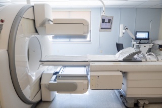
PET (positron emission tomography) scans provide details on how well your organs and tissues are working. Injected through an IV, the tracer used for this test absorbs into your body and helps the PET scanner take 3D pictures. PET scans take about two to three hours to complete.
SPECT (single-photon emission computerized tomography) shows your body’s functional processes, such as blood flow to your heart or brain, and helps your doctor detect any abnormalities. The injected tracer may take several hours for your body to absorb before the scanning of your body and area of concern can begin.
A bone scan uses a special type of radioactive tracer that emits radiation as it collects in your bones. After the tracer is injected, you may have to wait up to three hours before the scan can be started. Some exams, such as three-phase bone scans, may be broken up into two visits with four hours in-between each scanning session.
A cardiac scan uses a radioactive tracer that emits energy in your heart, where it helps the scanner create 3D pictures of this organ. Cardiac scans are typically done before and after you have completed an exercise stress test on a treadmill or stationary bike. From start to finish, the cardiac stress test and scanning takes about three and a half hours.
HIDA scans, or hepatobiliary scans, involve radioactive tracers that travel from your liver into your gallbladder and small intestine. After the tracer is injected into your body, a scanner tracks it as it moves through your digestive system. HIDA scans help diagnose liver or gallbladder disease. The test lasts about one and a half hours.
View Your Radiology Images
MyChart is your personalized patient portal and keeps you connected to your imaging and radiology records — no matter where you are.

Awards and Certifications
Nuclear Medicine Technologist Certification Board
UVM Health technologists who perform nuclear medicine procedures are certified by the Nuclear Medicine Technologist Certification Board, meaning they have met the rigorous standards of the specialty. Ongoing continuing medical education ensures our technologists are kept up to date on the latest nuclear medicine imaging techniques and therapeutic approaches to provide you with the safest and highest level of care.
Locations near you
Share your location to see nearby providers and availability
62 Tilley Drive
South Burlington, VT 05403
111 Colchester Avenue
Main Campus, McClure, Level 1
Burlington, VT 05401
75 Beekman Street
2nd Floor
Plattsburgh, NY 12901
111 Colchester Avenue
Main Campus, McClure, Level 1
Burlington, VT 05401-1473
133 Park Street
Malone, NY 12953-1241
130 Fisher Road
Berlin, VT 05602-9516
115 Porter Drive
Middlebury, VT 05753-8423