Bone Densitometry
Bone Densitometry at UVM Health
A commonly used screening and diagnostic test, bone densitometry measures bone loss to determine the health of your bones and risk for fractures. Also called DEXA or DXA (dual-energy X-ray absorptiometry), bone density scans use very small doses of ionizing radiation to capture X-ray images of your body’s skeleton, usually focusing on the hips and lower spine, where many fractures occur.
Quick and noninvasive, bone density scans help reveal if you have brittle, thinning bones. These can occur for many reasons, from your age and diet to disease. Bone densitometry is a painless standard imaging technique, and is the most frequently used method for diagnosing osteoporosis, a condition that often affects women after menopause but may also be found in men.
Why Choose UVM Health?
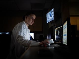
At University of Vermont Health, our radiologists have extensive experience in analyzing DEXA images to efficiently deliver timely and accurate results. We provide your doctor with key information about your bone mass to aid diagnosis, treatment and care management for a variety of medical conditions.
Highlights of our DEXA services include:
- Skilled bone densitometry team: With your comfort a top priority, our densitometric technologists quickly work to capture high-quality images for the best assessment of your bone density.
- State-of-the-art technology: We use the latest bone densitometer systems available, which provide superb imaging resolution and take precise measurements for the most accurate results. Using low-dose X-rays, they reduce your radiation exposure to the safest levels possible.
- Experienced, trusted expertise: Our in-demand DEXA services deliver quality results to guide your provider in evaluating and treating osteoporosis and other bone diseases. We offer convenient access throughout UVM Health to meet the diagnostic screening and health-monitoring needs of the communities we serve.
Common Uses for Bone Densitometry
Bone loss often develops slowly and silently over time, with few noticeable symptoms. Bone density testing is an easy, painless way to diagnose, and track the benefits of, treatment for a wide array of health problems, such as:
- Arthritis
- Bone cancers
- Bone pain
- Cancer metastasis to the bones
- Fractures
- Infections
- Osteoporosis
Lifestyle choices such as smoking or excessive alcohol consumption can also promote bone loss.
What to Expect During a Bone Density Scan
Bone densitometry exams require very little advance preparation. On the day of your DEXA, you may eat normally, but you should avoid taking calcium supplements for at least 24 hours before the procedure.
For an examination of your hip(s) or spine, you will lie on your back on a padded table. An X-ray generator will be located below you and an imaging device, or detector, will be positioned above you. To assess your spine, one of our specially trained technologists will place a padded positioning block under your legs to help flatten your pelvis and lower (lumbar) spine to ensure we capture the best images. To assess your hip, you may need to place your foot on a brace that rotates your hip inward. In both cases, the detector will slowly pass over the area of your body being scanned.
To avoid blurred images, you must hold very still and may need to hold your breath for a few seconds during the scanning. The technologist will walk behind a wall or into the next room to activate the X-ray machine.
Exams of the finger, hand, forearm or foot are simpler. You will place the body part being examined into a small bone densitometry device. It generates a reading of your bone mass within a few minutes.
View Your Radiology Images
MyChart is your personalized patient portal and keeps you connected to your imaging and radiology records — no matter where you are.

Locations near you
Share your location to see nearby providers and availability
101 Adirondack Drive
Ticonderoga, NY 12883
75 Park Street
Elizabethtown, NY 12932
89 Plaza Boulevard
Plattsburgh, NY 12901
192 Tilley Drive
South Burlington, VT 05403-4440
1311 Barre Montpelier Road
Suite 400
Berlin, VT 05602
133 Park Street
Malone, NY 12953-1241
130 Fisher Road
Berlin, VT 05602-9516
115 Porter Drive
Middlebury, VT 05753-8423
76 McNeil Road
Suite 2
Waterbury Center, VT 05677-7162