Diabetic Retinopathy
Diabetic Retinopathy Care at UVM Health
Diabetic retinopathy occurs when high blood sugar levels from diabetes damage blood vessels in your eye’s retina. Your retinas send signals from your eyes that your brain uses to make images and are crucial for sight. Diabetic retinopathy often affects both eyes and is the top cause of blindness in adults younger than 65.
We know the idea of losing your vision is alarming. University of Vermont Health ophthalmology program is part of an academic medical center. Our retina specialists not only offer the latest treatments to protect your vision, they are also active in research to improve eye disease care.
Why Choose UVM Health?
As one of the leading comprehensive ophthalmology programs in the region, we offer:
- Dedicated retina specialists: You receive eye care from eye doctors (ophthalmologists) who have advanced fellowship training and board certifications in retinal diseases. These specialists have decades of experience with diabetic retinopathy.
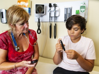
- Team approach to care: High blood sugar levels can worsen diabetic retinopathy and speed vision loss. We partner with UVM Health endocrinologists and primary care providers to promote proper diabetes management that protects your vision.
- Advanced surgical treatments: We always start with the least invasive therapies. Should you need more advanced care, we have the expertise to perform complex eye surgeries to save your eyesight.
Types of Diabetic Retinopathy
There are two stages, or types, of diabetic retinopathy:
- Nonproliferative diabetic retinopathy (NPDR): During the early stage of disease, blood vessels in your retina leak. This leakage causes swelling (macular edema). Blood vessels may also close off, stopping blood from reaching your retina (macular ischemia).
- Proliferative diabetic retinopathy (PDR): In this advanced disease stage, the retina grows new blood vessels that leak blood into your eyes. This can cause retinal detachment.
Symptoms of Diabetic Retinopathy
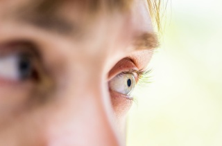
Early-stage diabetic retinopathy rarely causes symptoms. As more eye damage occurs, you may experience:
- Changes in how you see colors
- Distorted or blurry vision
- Eye floaters (dark spots or streaks in your vision)
- Poor night vision
- Trouble reading
I'm grateful for the excellent care I received which resolved significant problems I had with one eye. They paid close attention to my situation following my operation. I feel fortunate to have had such a knowledgeable and concerned team.
Diagnosing Diabetic Retinopathy
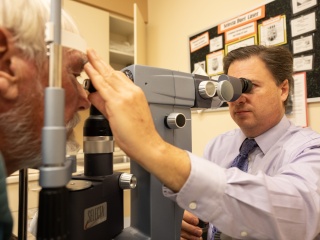
Our retina specialists will review your symptoms and medical history. You may also get one or more of these diagnostic tests:
- Eye examination: You’ll undergo vision tests and a dilated eye exam to help your provider look inside your eye.
- Optical coherence tomography (OCT): This noninvasive 3D retinal imaging scan measures light reflecting off your retina. It can show retinal swelling.
- OCT angiography (OCT-A): This advanced OCT scan uses the reflections of laser lights on blood to create 3D images of retinal blood flow.
- Fluorescein angiography: Your provider uses a special camera to view an injectable dye as it moves through blood vessels in your retinas.
Diabetic Retinopathy Treatment
We offer a full range of nonsurgical and surgical treatments for diabetic retinopathy. We always start with the least invasive treatment first.
Injections of anti-vascular endothelial growth factor (anti-VEGF) medication into your eyes can stop the production of VEGF. This protein causes leakage and helps form new blood vessels. Anti-VEGF injections can slow vision loss and improve your sight.
Laser photocoagulation uses a laser to help shrink blood vessels and keep them from growing. We also use laser therapy to help stop retinal swelling.
Our retina specialists have fellowship training in vitrectomy surgery to treat advanced diabetic retinopathy. During this outpatient procedure, your provider removes some or all of the vitreous gel from the back of your eye. This clear fluid helps maintain the shape of your eye and allows light to pass through to the retina.
Your provider may replace the gel with a saline (salt water) solution or a bubble of gas or oil. Over time, your eye makes more aqueous humor (a clear liquid in the front of the eye) to replace the saline or bubble. Your provider also removes blood and scar tissue around the retina. This treatment makes it easier for light rays to reach your retinas.
Locations Near You
Share your location to see nearby providers and availability
58 East View Lane
Berlin, VT 05641-5324
350 Tilley Drive
Suite 101
South Burlington, VT 05403-4539